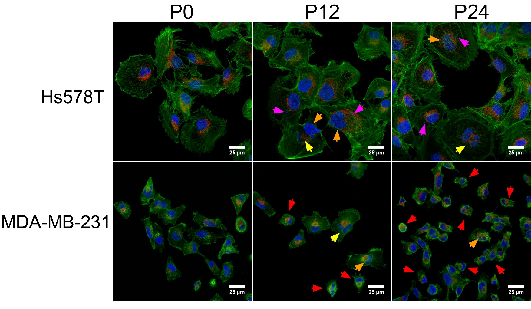
Fig. 2. Alterations of cellular morphology after a prolonged exposure with multiple doses of 0.1 nM paclitaxel. Morphological traits were visualized using confocal microscopy. The cellular components were stained with Phalloidin-FITC dye specific for actin filaments, DAPI dye for nucleus and MytoTracker for mitochondria. Scale represents 25 µm. Images were captured with 60x oil immersed objective. Magenta arrows represent the formation of the micronucleus, yellow arrows point multinucleate/polynuclear cells, red arrows denote cellular shrinkage and orange arrows point nuclear damage/fragmentation.